
Figure 3.
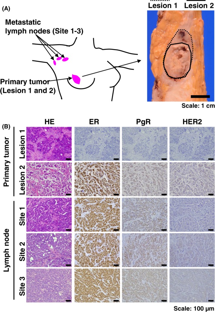
Breast cancer patient with different histological subtypes in primary and metastatic sites. (A) Pink circles indicate primary breast cancer and lymph node metastatic sites at levels I and II. Macroscopic image of corresponding primary tumor is shown on the right. In the primary tumor, the solid line indicates the hormone receptor (HR) (‐) and HER2 (‐) site in lesion 1 and the dotted line indicates the HR (+) and HER2 (‐) site in lesion 2. Scale bar: 1 cm. (B) Representative image of HE and immunohistochemical staining. Primary tumor at lesion 1 indicates the estrogen receptor (ER)− PgR− HER2−, tumor at lesion 2 indicates ER + PgR+ HER2−, and all the metastatic lymph nodes showed ER − PgR− HER2−. Scale bar: 100 μm.