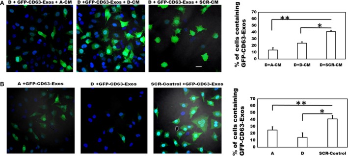
Figure 6.

Fetuin‐A promotes the uptake of GFP‐CD63‐labeled exosomes in LN229 subclones. In (A), fetuin‐A knockdown subclone D cells were seeded at 1 × 10⁴ cells/well in three of the wells of a 4‐well glass slide. After an 8 h incubation in SFM, the medium in the wells was replaced with previously collected conditioned medium (1 mg/mL; 200 μL/well) from fetuin‐A knockdown subclone (A) (A‐CM); subclone (D) (D‐CM), and SCR‐Control (SCR‐CM) cells. After setting the GFP‐channel to the lowest threshold, labeled exosomes (isolated and purified from BT‐CD63; 20 μg/mL) were then added to each well (30 μL/well) and incubated for 1 h. The cells were fixed in 4% formalin (w/v), a drop of slow fade with DAPI added to each slide, coverslipped, and examined under a Nikon A1R confocal microscope. The number of green cells (containing exosomes) in a 20× microscopic field (five different fields/data point), were counted and expressed as percentage of total cells (green+blue) per field. The bars represent mean ± SEM of two separate experiments (*P = 0.0003; **P = 0.0001). In (B), the LN229 subclones (A, D, and SCR‐Control) were seeded in three of the wells of a 4‐well glass slide as above in SFM and incubated for 8 h. After adjusting the GFP‐fluorescence channel to the lowest threshold as in (A) above, the GFP‐labeled exosomes (20 μg/mL) were added to each of the three wells and incubated for 1 h, cells fixed in 4% formalin, and prepared as above for confocal microscopy. The percentage of green cells was determined as above and the bars represent mean ± SEM of two separate experiments (*P = 0.0002; **P = 0.0004; scale bar represents 10 μm). SFM, serum‐free medium.
