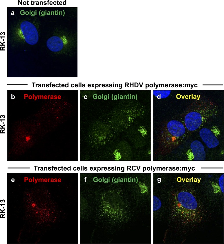
Fig 2. Redistribution of the cis/medial Golgi membrane marker giantin in cells expressing rabbit calicivirus RdRps.
RK-13 cells were transiently transfected with expression constructs coding for myc-tagged versions of RHDV RdRp (b–d) or RCV RdRp (e–g). Cells were fixed 24 h after transfections and recombinant proteins were immunostained using anti-myc antibodies (shown in red) and anti-giantin antibodies as a Golgi membrane marker (shown in green). DAPI was used to stain cell nuclei (shown in blue). In cells expressing RHDV or RCV RdRps (b and e, respectively), a striking redistribution of the Golgi membrane marker was observed (c–d and f–g, respectively) as compared with the normal Golgi structure in untransfected control cells (a).

