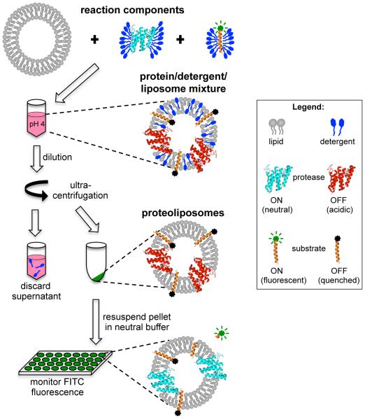
Figure 1.
Schematic diagram of the inducible reconstitution and fluorogenic intramembrane protease assay. Pure rhomboid protease and FITC-TatA substrate in detergent micelles are mixed with liposomes at low pH to inactivate the protease during reconstitution. Detergent is removed from the protein/lipid/detergent complexes by means of dilution followed by ultracentrifugation. Resuspension of the proteoliposome pellet in neutral reaction buffer re-activates the protease. The FITC fluorophore on the reconstituted substrate is quenched by its proximity to the membrane lipids, allowing detection of a fluorogenic signal as evidence of proteolytic release of the FITC-labeled amino-terminus.

