. 2016 Dec 22;5:e20894. doi: 10.7554/eLife.20894

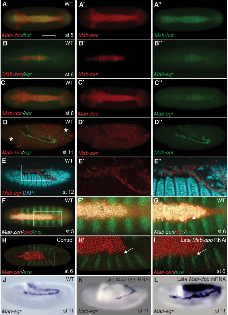
Figure 2. Specification of amnion by BMP signaling in Megaselia.

(A) Mab-hnt and Mab-doc expression at the late blastoderm stage. Scale bar = 100 µm. (B, C) Mab-egr and Mab-zen (B) and Mab-egr and Mab-doc (C) expression at early gastrulation. (D) Mab-egr and Mab-zen expression after germ band extension. Asterisks denote tears in the serosa during sample preparation. (E) Mab-egr expression during germ band retraction. The serosa has been removed and nuclei have been labeled with DAPI. Boxed region enlarged (E’–E’’). (F, G) Mab-doc, Mab-zen and Mab-eve expression at late blastoderm stage (stage 5) (F, enlargement F’) and early gastrulation (stage 6) (G) with arrow pointing to abutting Mab-eve and Mab-doc expression domains. (H, I) Mab-zen and Mab-eve expression in early gastrula control embryo (H, enlargement H’) and following Mab-dpp knockdown after 50% blastoderm cellularization (I). Arrows, gap between the Mab-eve and Mab-zen domains (H’) that is suppressed in the knockdown embryo (I). (JL) Mab-egr expression at germ band extension in wild-type embryo (J), and after Mab-dpp knockdown (K) or Mab-dpp overexpression (L) after 50% blastoderm cellularization.

DOI: http://dx.doi.org/10.7554/eLife.20894.003

Figure 2.

Figure 2—figure supplement 1. Expression of Mab-doc2.

Figure 2—figure supplement 1.

(A, B) Mab-doc2 expression at early gastrulation (A) and the extended germ band stage (B). (C) Maximum likelihood gene tree based on full-length Doc protein homologues. Aae (Aedes aegypti), Aga (Anopheles gambiae), Mab (Megaselia abdita), Dme (Drosophila melanogaster), Dps (Drosophila pseudoobscura), Dgr (Drosophila grimshawi). Bootstrap values, based on 1000 replicas, are shown. (A) Dorsal views with anterior left. (B) Lateral views with dorsal up and anterior left.
Figure 2—figure supplement 2. Time course of Mab-zen, Mab-doc and Mab-eve expression.

Figure 2—figure supplement 2.

(AC) Mab-zen, Mab-doc and Mab-eve expression in Megaselia embryos with the second panel showing enlarged view near the posterior Mab-eve strips, followed by single channel images of DAPI, Mab-eve, Mab-doc, and Mab-zen in successive panels at early blastoderm (A), late blastoderm (B) and early gastrulation (C) stages, indicating amnion specification occurs at early gastrulation. Dorsal views with anterior left.
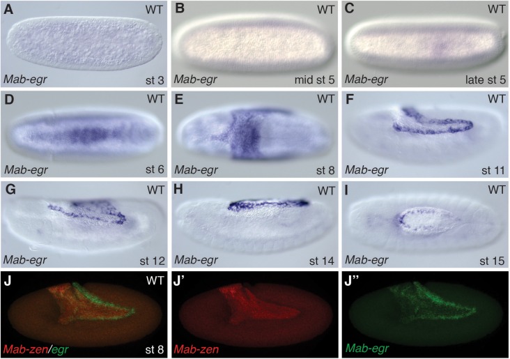
Figure 2—figure supplement 3. .Expression of Mab-egr.

Figure 2—figure supplement 3.

(AI) Mab-egr expression in Megaselia embryos before blastoderm formation (A), during blastoderm cellularization (B), and at cellular blastoderm (C), early gastrulation (D), early and late germ band extension (E, F), germ band retraction (G) and dorsal closure stages (H, I). (J) Mab-egr and Mab-zen expression in embryos during germ band extension. (AE, I) Dorsal views with anterior left. (FH, J) Lateral views with dorsal up and anterior left.
Figure 2—figure supplement 4. Overexpression of Mab-eve represses Mab-zen expression.

Figure 2—figure supplement 4.

(A, B) Two examples of Mab-zen and Mab-eve expression at early gastrulation after Mab-eve overexpression. Dorsal views with anterior left.