Fig. 4.
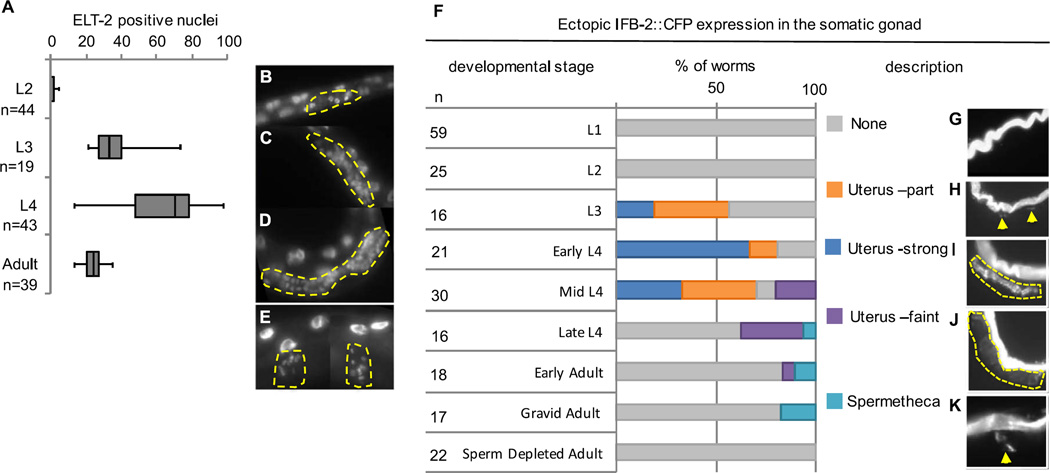
Organogenesis of the somatic gonad can be redirected into intestine at both proliferative and post-mitotic stages. (A) Comparison of the number of immunoreactive ELT-2 nuclei in the somatic gonad after ectopic ELT-7 expression at the indicated stage. n, number of worms. ELT-2-expressing nuclei in the proximal gonad (yellow outline) 48 h after pulsed ELT-7 expression at the L2 (B), L3 (C), and L4 (D) stage. (E) ELT-2-expressing nuclei in the spermatheca (yellow outline) after ectopic ELT-7 expression at the adult stage. (F) Percentage of worms with IFB-2::CFP expression in the somatic gonad 48 h after pulsed ELT-7 expression at the indicated stages determined by worm length and vulval morphology. n, number of worms. (G–H) Typical example of the described phenotypes with yellow arrows and dotted lines demarcating the region of ectopic IFB-2::CFP expression. (G, none) no ectopic IFB-2. (H, Uterus-part) Some ectopic IFB-2 that does not form a complete lumen. (I, Uterus-strong) IFB-2 expression similar to intestine that outlines an intestine-like lumen. (J, Uterus-faint) Faint IFB-2 expression that outlines a wider more uterus-like lumen; (K, Spermathecae) ectopic IFB-2 in one or both spermathecae.

