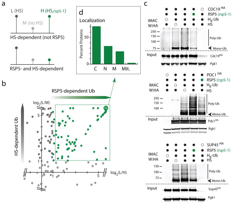
Figure 3. Rsp5 targets mainly cytosolic proteins upon HS.
a. Schematic representation of the triple SILAC experiment to identify HS-induced Rsp5 substrates (L:light; M:medium; and H: high labels). b. Plot of the log2 ratios of each quantified ubiquitinated peptide. In the y-axis, ratios of L versus M are reported to display sites affected by HS, and ratios of L versus H are reported in the x-axis to indicate sites affected by the absence of Rsp5 activity. RSP5-dependent ubiquitination sites are indicated in green. The are 76 overlapping sites with log2(ratios) ≥ 5.64. c. IMAC-enriched ubiquitin conjugates were analyzed by western blots. Samples were derived from indicated cells (rsp5-1 designated by green circles) was assed expressing endogenously tagged candidates (3xHA) and plasmid version of H8-Ubiquitin. All uncropped images are in Supplementary Figure 8. d. Localization of HS-induced Rsp5 substrates identified in b (C: Cytoplasm; N: Nuclear; M: Membrane; Mit: Mitochondria; source values are listed in Supplementary Table 5).

