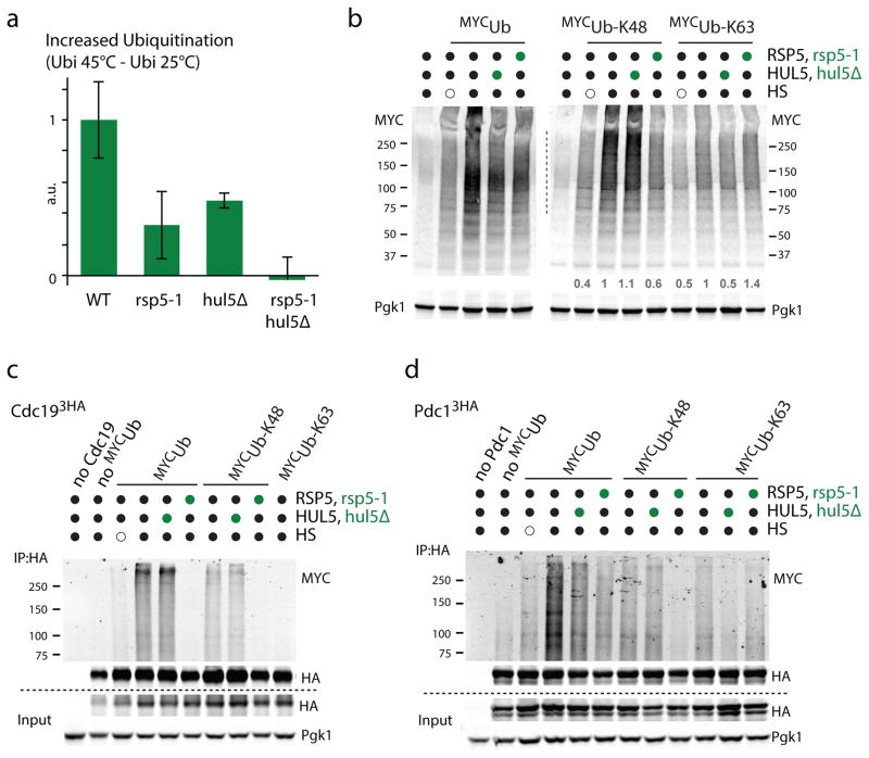
Figure 4. Both Rsp5 and Hul5 mostly target their substrates independently.
a. Increased ubiquitination levels quantified by dot-blot after a 15min HS at 45°C in the indicated cells. Experiment was done with three biological replicates (average±SD; source values are listed in Supplementary Table 5) and a.u. denotes arbitrary units. b. Western blot analysis of the indicated cells that expressed on a plasmid the indicated N-terminally MYC tagged ubiquitin constructs (K48 and K63 designate ubiquitin variants that only contain K48 and K63, respectively, while all other lysines are mutated to arginines) that were subjected or not to HS (45°C, 15min). c–d. C-terminally 3xHA tagged Cdc19 (c) and Pdc1 (d) expressed from their endogenous promoter on a plasmid were immunoprecipitated from the indicated cells (hul5Δ and rsp5-1 designated by green circles) expressing the indicated N-terminally MYC tagged ubiquitin constructs. Cells were HS or not for 20min at 45°C. Wild-type MYC tagged ubiquitin (MYCUb) was also expressed in the control cells used in lane 1. All uncropped images are in Supplementary Figure 8.

