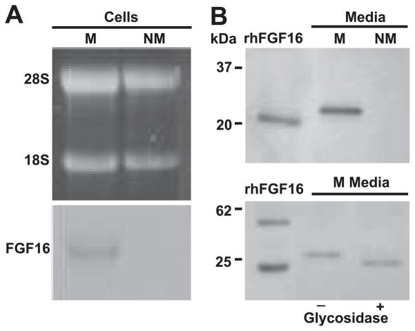
Fig. 2.
FGF-16 is produced by neonatal cardiac myocytes in culture. A: total RNA (40 μg) isolated from cell cultures enriched for either myocytes (M) or nonmyocytes (NM) was visualized with ethidium bromide (top) and then blotted and probed for FGF-16 transcripts. A 1.8-kb FGF-16 transcript (relative to the 28S and 18S ribosomal RNA bands) was detected in myocytes but not nonmyocytes. B, top: neonatal rat cardiac cell cultures enriched for myocytes and nonmyocytes were processed and conditioned media from each cell type were enriched for heparin binding proteins. Samples were separated by SDS-PAGE and probed using custom polyclonal FGF-16 antiserum. A band of 26.5 kDa was seen in myocytes but not nonmyocytes. (B, bottom). Conditioned media were incubated with glycosidase buffer in the absence (−) and presence (+) of enzyme. Fractions were separated by SDS-PAGE and probed with commercial FGF-16 antibody. Recombinant human (rh) FGF-16 (10 ng) and mobilities of markers (B, left) are included as specificity or size controls, respectively. B, bottom, left lane: rhFGF-16 appears as both a monomer and a dimer.

