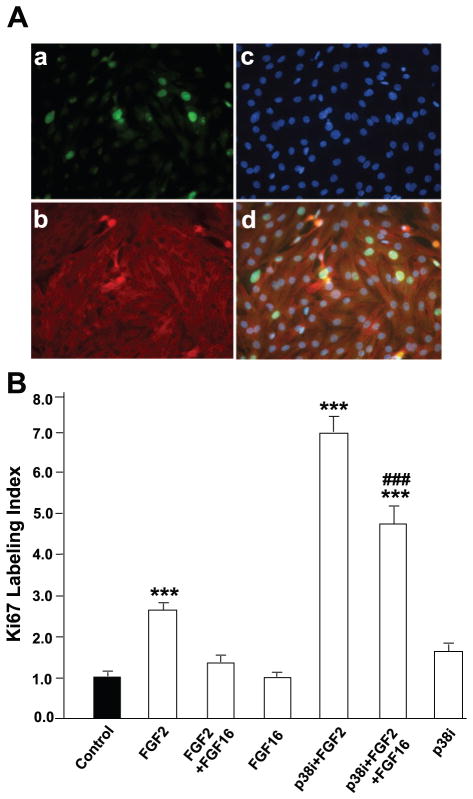
Fig. 3.
FGF-16 interferes with FGF-2-induced Ki67 labeling index in neonatal rat cardiomyocytes. A: cells cultured on coverslips were treated with 1 ng/ml FGF-2 and 100 ng/ml FGF-16 in the presence or absence of 10 μM p38 inhibitor (p38i, SB203580) for 24 h, fixed, and stained simultaneously with anti-Ki67 antibody (a), α-actinin (b), and Hoechst 33342 staining for detection of nuclei (c); d is merged image of a, b, and c. In this manner, the Ki67 labeling index was determined as the fraction (percentage) of Ki67-positive myocytes (identified by α-actinin staining). B: labeling index was counted in 59–67 fields for each treatment group, and value for control (untreated) cells was set to 1.0. Absolute means ± SE for control group (n = 67) are 1.68 ± 0.24%. ***P < 0.001, compared with control; ###P < 0.001, compared with FGF-2 treatment in the presence of p38 inhibitor, using one-way ANOVA and Tukey-Kramer posttest for multiple comparisons.

