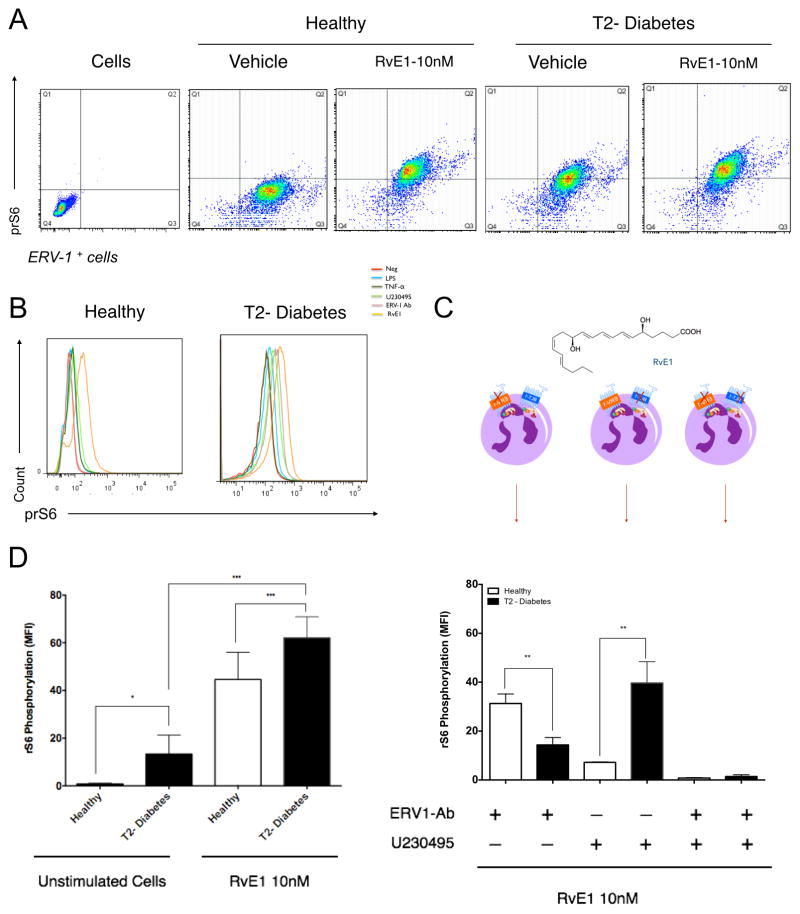
Figure 3. Signaling by rS6 phosphorylation is regulated through RvE1/ERV1 axis.
Activation of ERV1 receptor by RvE1 is through rS6 signaling. (A) RvE1 induces rS6 phosphorylation of healthy and diabetic neutrophils as demonstrated by gating ERV-1 positive cells (10nM, 30min). (B) To quantify phospho-rS6 expression of healthy and diabetic populations, unstimulated cells were evaluated and compared to RvE1 treated cells. (C) Phosphorylation of rS6 was measured after blocking ERV1 and BLT1 receptors. Cells were treated with RvE1 (10nM) alone, or in combination with BLT1 receptor antagonist (U-230495) or ERV1 receptor antagonist (ERV1/Ab) or both. (D) Cells were treated with various stimuli to investigate the specificity of rS6 phosphorylation LPS (10ng/ml), TNFα (10ng/ml), U-230495 (10nM), ERV1/Ab (10ng/ml), RvE1 (10nM). Results are expressed as mean fluorescence intensity (MFI, mean ± SEM). Statistical significance was evaluated by Wilcoxon test (n=16; healthy n=8, T2D, n=8, * p<0.05, ** p<0.01, *** p<0.001, ns = non-significant).

