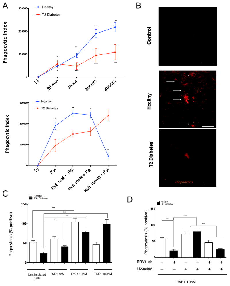
Figure 6. Neutrophil phagocytosis is mediated by ERV-1 activation.
Phagocytosis rate analysis of Porphyromonas gingivalis (Pg) and zymosan particles by peripheral blood neutrophils obtained from healthy and type 2 diabetic adult volunteers. Pg was labeled with BacLight and opsonized in heat inactivated normal serum 30 minutes at room temperature and incubated with neutrophils from both groups (MOI = 20:1). (A) The rate of phagocytosis of positive labeled bacteria was evaluated by flow cytometry. (B) Deep red labeled zymosan particles were incubated with neutrophils (1 hour) and evaluated by immunofluorescence microscopy. (C) The actions of RVE1 treatment (1–10 nM) on phagocytosis were quantified. (D) Labeled bioparticles were readily visible within neutrophils and expressed as mean ± SD mean fluorescence intensity. Phagocytic index was measured after blocking ERV1 and BLT1 receptors. Cells were treated with RvE1 (10nM) alone, or in combination with BLT1 receptor antagonist (U-230495) or ERV1 receptor antagonist (ERV1/Ab) or both. Phagocytosis experiments are expressed as phagocytic index (% positive x MFI). Statistical significance was evaluated by Wilcoxon test (n=24; healthy n=12, T2D, n=12, * p<0.05, ** p<0.01, *** p<0.001, ns = non-significant).

