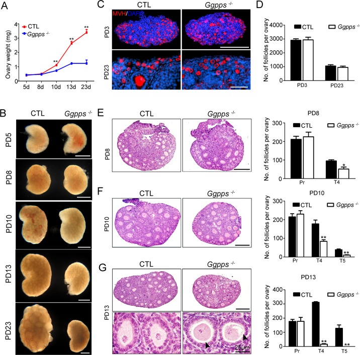
Fig 3. GGPP depletion in oocytes inhibits ovarian primary-secondary follicle transition.
(A) Ovary weight at the indicated time points (n = 3–6). (B) The images of the ovaries at different time points were captured using a light microscope. (C and D) MVH immunofluorescence and primordial follicle numbers (n = 4) in PD 3 and PD 23 ovaries from Ggppsfl/fl Ddx4-Cre and CTL mice. DAPI (blue) indicates the cell nuclei. (E-G) H&E staining of ovaries at the indicated time points and quantification of the different types of follicles observed in ovaries from PD 8, PD 10, and PD 13 mice, including primordial (Pri), primary (Pr), type 4 (T4), and type 5 (T5) follicles. The number of follicles per ovary was quantified as described in Materials and Methods (n = 4). Arrowheads indicate abnormal contact between the oocyte and granulosa cells. Data were presented as the mean ± SEM. * p<0.05, **p<0.01. Scale bar, 200 μm.

