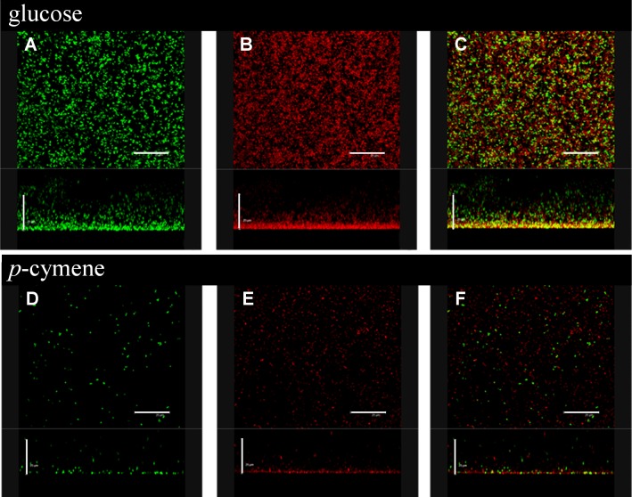
Fig 7. Effect of p-cymene on biofilm formation by B. xenovorans LB400.
Cells were grown in M9 medium using glucose or p-cymene as sole carbon source until exponential phase (Turbidity600nm = 0.6) and washed were resuspended in M9 medium supplemented with glucose (5 mM) or p-cymene (vapor phase). Microscopic characterization of B. xenovorans LB400 biofilms grown in M9 medium supplemented with glucose (5 mM) or p-cymene (vapor phase) during 48 h. Cells in the biofilms were stained with the BacLight kit showing viable (green fluorescence) and non-viable (red fluorescence) bacteria (C and F). The viable and non-viable cells were marked with green fluorescence (A and D). The dead cells were stained red (B and E). Images were horizontal and vertical three-dimensional reconstructed in the x–y plane and x–y plane, respectively. In all images the scale bar is 25 μm.

