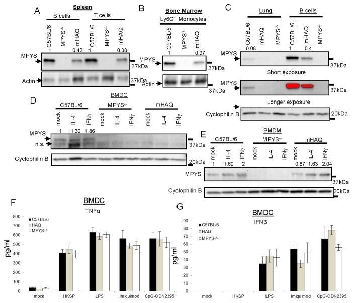
Figure 3. The HAQ knock-in mouse (mHAQ) has decreased MPYS expression in multiple tissues.
A–C. Various types of cells from the C57BL/6, HAQ, and MPYS−/− mice were lysed in RIPA buffer and probed for indicated Abs as in Figure 1 (n=3). D–E. BMDC or BMDM from the C57BL/6, mHAQ, and MPYS−/− mice were treated with IL-4 (40ng/ml) or IFNγ (40ng/ml) or mock (PBS) overnight. Cells were lysed in RIPA buffer and probed for indicated Abs as in Figure 1 (n=3). F–G. BMDC from C57BL/6, MPYS−/− or mHAQ were stimulated with HKSP (107c.f.u/ml), LPS (20ng/ml), imiquimod (4ng/ml) or CpG-ODN2395 (8ng/ml) for 5hrs. TNFα (F) and IFNβ (G) were measured in the cell supernatant (n=3). Graph present means ± SEM from three independent experiments.

