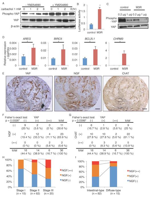
Figure 7. M3R activates YAP signaling in human gastric cancer cells.
(A) Immunoblotting of TMK-1 cells treated with 1 mM carbachol for the indicated times. Cells were pretreated with vehicle or 10 m YM254890. β-actin was used as a loading control. (B-D) Relative YAP luciferase activity (B, n = 3/group), immunoblotting (C), and relative gene expression (D, n = 3/group) in AGS cells transfected with control or M3R-expressing vectors. Samples are collected 24 hours after transfection. (E) Representative images of YAP, NGF, ChAT staining in human gastric cancers. (F-G) Correlation between the expression levels of YAP and NGF (F), and of YAP and ChAT (G) in 36 gastric cancer cases. (H-I) NGF positivity in different cancer stages (H) and histological forms (I) in 97 gastric cancer cases. Means ± SEM. *p < 0.05 (t-test in B and D, Fisher in F and G). Bars = 200 m. See also Figure S7, Table S1, and Table S2.

