Figure 1.
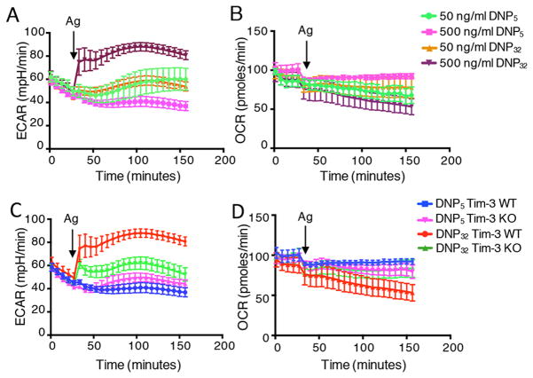
IgE/Ag-activated mast cells undergo a rapid increase in glycolysis. (A–B) WT BMMCs were sensitized with IgE for three hours and activated in-Seahorse with the indicated concentrations of DNP32 and DNP5 for two hours. Data in panel A show the change in glycolysis (ECAR), while panel B shows mitochondrial respiration (OCR). (C–D) WT and Tim-3 KO BMMCs were sensitized with IgE for three hours and stimulated with indicated antigens directly in-Seahorse for two hours. Arrow indicated when antigen was injected. Data are representative of two (E, F) and three independent (A–D) experiments.

