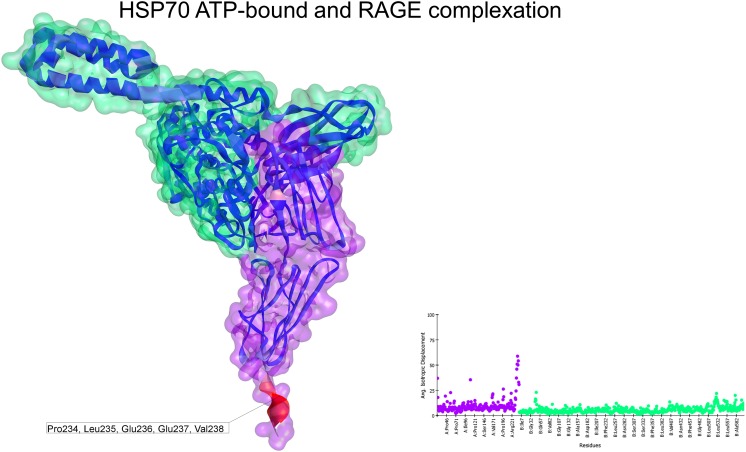
Fig. 5.
Normal mode analyses for the complex 3CJJ-4B9Q. Structures are shown according to their respective residue deformability. The color scale ranges from blue to red with increasing flexibility of residues. Every structure includes a corresponding scatter plot for residue deformability. There was a highly flexible region at the end of the C1 domain of the receptor and a moderately flexible region on the helical lid of HSP70. Overall, this complex exhibited a flattened profile of residue deformability and flexibility compared with the other complexes. Additionally, the interacting region was colored blue, indicating the relative stability of this complex. RAGE surface is colored purple and HSP70 surface is colored green. Residues that most contribute to the deformability of the system are indicated

