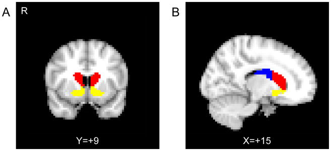
Figure 2.
Striatal subregion masks used in the analysis. A) The coronal plane anterior to the plane of the anterior commissure is shown, containing the ventral striatum (yellow) and the anterior caudate (red). B) The sagittal plane showing the division between the anterior caudate (red) and the posterior caudate (blue), as defined by the anterior commissure.

