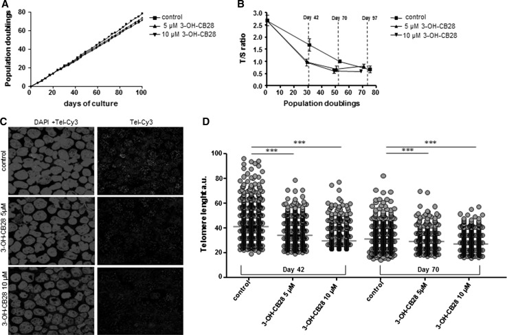
Fig. 5.
a Population doublings of cultured Jurkat T cells for 100 days of culture. Cells were cultured in the presence of 3-OH-CB28 at a concentration of 5 or 10 µM, or with solvent control. b Telomere length analysis of Jurkat T cells, cultured as described in a and analyzed by MM-qPCR. Relative TL is expressed as T/S ratio and plotted against population doublings. Days of culture (day 42, day 70, day 97) are indicated. c Representative images of cultured Jurkat T cells at day 42 of culture analyzed by confocal quantitative fluorescence in situ hybridization (confocal Q-FISH). Cells were cultured either with 3-OH-CB28 at a concentration of 5 or 10 µM, or with solvent control and subsequently stained with DAPI and a Cy3-labeled telomere probe on cytospin slides. d Quantification of confocal Q-FISH of Jurkat T cells by telomapping analysis for the indicated time points and culture conditions. TL quantification is given in arbitrary units of fluorescence (a.u.). Statistically significant differences are indicated (***p < 0.001)

