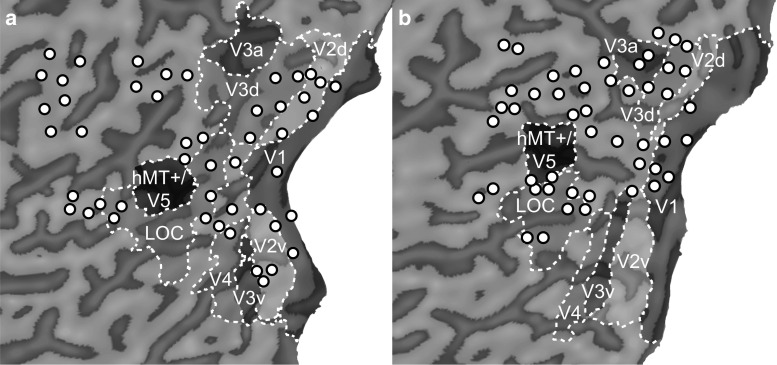
Fig. 4.
Flatmaps for two participants showing retinotopic areas, hMT+/V5 and LOC projected on the pial surface with Brainvoyager “VOI to POI plugin”. Sulci are shown in dark grey, gyri in light grey. For each stimulation target, we defined a centre of TMS-related effects (circles). Due to the decay of TMS-related effects over distance, they are mostly located on gyral crowns. Depending on individual brain anatomy, we were not able to target areas with TMS that are located on the bottom of a sulcus (a V3a and hMT+/V5) or buried between cerebrum and cerebellum (b V2v, V3v, and V4)

