Fig. 1.
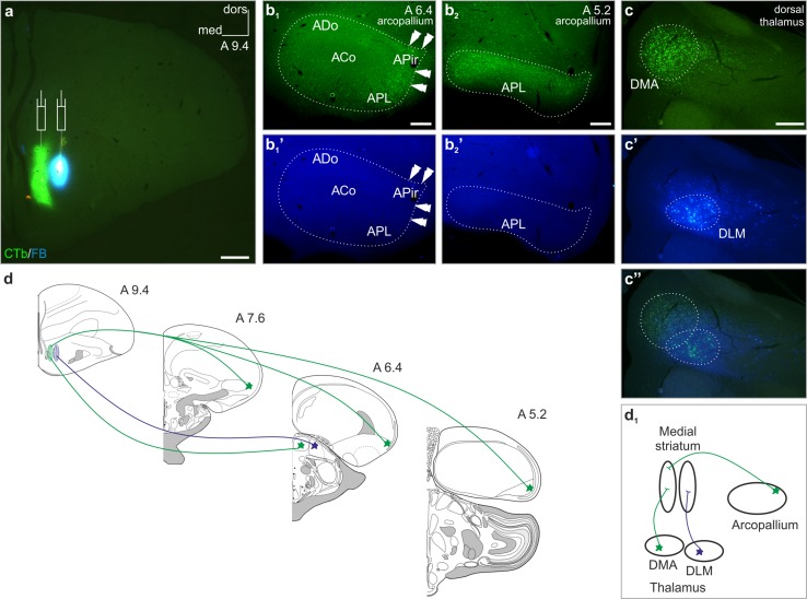
The medial striatum is inhomogeneous in its afferentation pattern. Simultaneous injections (appropriate symbols in a) of Alexa Fluor® 488 conjugated choleratoxin B subunit (CTb) and Fast Blue (FB) retrograde tracers into the medial and lateral divisions of the medial striatum, respectively. b 1, b 2 CTb-labeled (CTb+) somata (arrowheads) were detected only in the peripheral part of arcopallium, including the dorsal and posterolateral part of the arcopallium (ADo and APL, respectively), with an outstanding density in its lateral region previously termed as the amygdalopiriform area (APir) (Puelles et al. 2007). b 1 ′, b 2 ′ The arcopallium remained spared from FB-labeled (FB+) cell bodies. c–c′′ The striatal projection of the dorsal thalamus showed a medial–lateral topology with a narrow overlap between these territories: CTb+ perikarya were restricted to the anterior dorsomedial nucleus (DMA), whilst FB+ cell bodies to the medial part of the dorsolateral anterior thalamic nucleus (DLM). d, d 1 Schemata demonstrating the dual character of the retrogradely traced afferentation of the medial striatum (coronal section drawings were modified after the templates: http://www.avianbrain.org/nomen/Chicken_Atlas.html. Numbers at the right upper corner of the drawings and images indicate distance in millimeters AP according to Kuenzel and Masson (1988)). White dotted lines mark the outlines of arcopallium. ACo arcopallial core, dors dorsal, med medial. Scale bars 1 mm (a), 200 µm (b 1, b 2, c)

