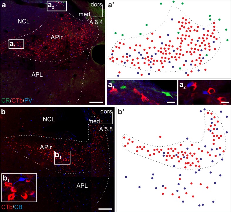
Fig. 5.
Arcopallial neurons projecting to BSTL and adjacent Ac are immunonegative for the major calcium binding proteins parvalbumin, calbindin and calretinin. (a, a′, a 1, a 2) The amygdalopiriform (APir) area of the arcopallium harbors a plethora of CTb+ neurons labeled retrogradely from the BSTL and adjacent Ac. These projection neurons do not express the calcium binding proteins calretinin or parvalbumin. Illustration (a′) shows the distribution pattern of single labeled calretinin+ (green circles), CTb+ (red circles) and parvalbumin+ (blue circles) neurons. (b, b′, b 1) Similarly, retrogradely labeled CTb+ neurons remained immunonegative for the calcium binding protein calbindin in a more caudal part of the same arcopallial region. Illustration (b′) shows the distribution pattern of single labeled CTb+ (red circles) and calbindin+ (blue circles) neurons. (a, b) Cranio-caudal levels of the coronal sections are indicated as distance in millimeters AP according to Kuenzel and Masson (1988). APL posterolateral amygdala, CB calbindin, CR calretinin, CTb choleratoxin B subunit, dors dorsal, med medial, NCL caudolateral nidopallium, PV parvalbumin. Scale bars 200 µm (a, b), 10 µm (a 1, a 2)

