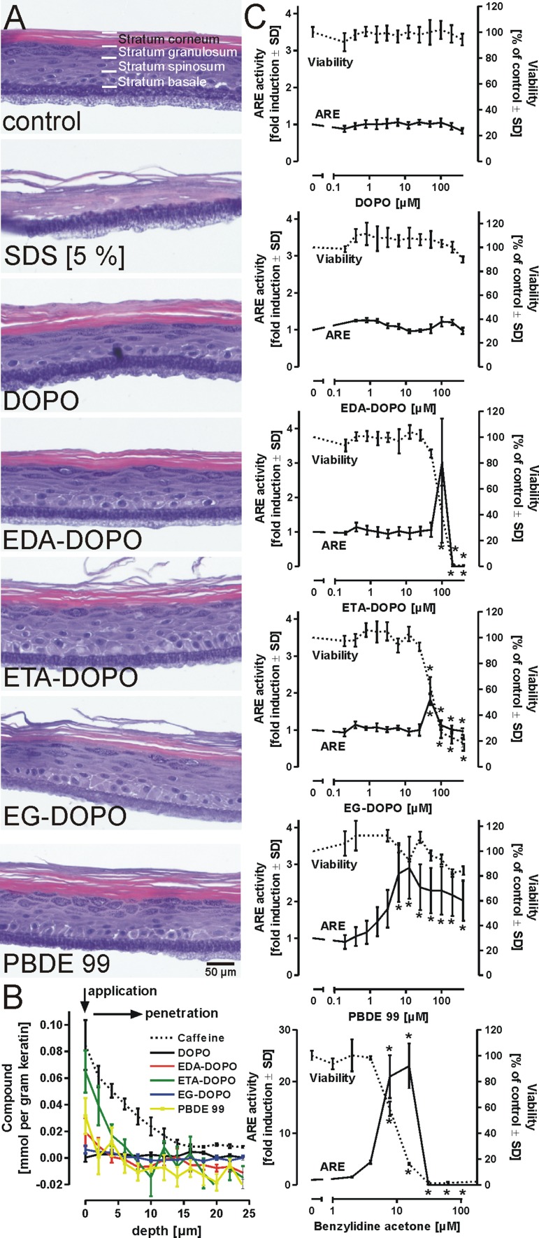
Fig. 3.
Interaction of DOPO derivatives with epidermal barriers. a Influence of flame retardants on epidermal integrity. The air-exposed stratum corneum of a 3D epidermal in vitro model generated from primary human keratinocytes was topically treated with various flame retardants (200 mM) for 15 min. The models were washed and further cultivated for 48 h. The tissues were analyzed by a viability assay and then fixed, sliced, and stained with hematoxylin and eosin to allow discrimination of epidermal layers. The loose layer beneath the stratum basale represents the filter membrane. b Penetration of flame retardants into skin. For the assessment of quantitative penetration profiles, porcine skin was topically exposed to the flame retardants (400 µM) for 1 h. Confocal Raman microscopy was applied to obtain a depth profile for each compound. The graph illustrates the amount of each flame retardant and that of positive control compound caffeine relative to keratin content as a function of depth. c Keratinocyte sensitization. To determine the skin sensitization potential of the tested flame retardants, KeratinoSens™ cells were stimulated with different concentrations of DOPO derivatives. Luciferase activity was detected 48 h after treatment, and the fold induction was calculated. Benzylidene acetone served as a positive control treatment. In parallel, the viability of the cells was assessed using the PrestoBlue® reduction assay. All data are mean ± SD (n = 3); *p < 0.05

