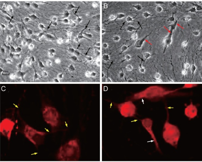
Figure 3. Morphological changes at the end of induction after Vitamin A for 24h.

A, B: Phase-contrast microscopic pictures (×200); A: Some EN orbital ADSCs showed synapsis-like structure (the black arrow) with evidence of cell-to-cell contact; B: Besides synapsis-like structure (the black arrow), a few EX orbital ADSCs exhibited tubular, rod-like structure (the red arrow) resembling outer segment of rod photoreceptors; C, D: Synapse and rhodopsin staining by immunofluorescence (IMF) staining. These images show synapse structure and rhodopsin expression (at the end of induction after vitamin A for 24h); C: EN orbital ADSCs. The red fluorescence is rhodopsin expression. Some cells showed synapsis-like structure (the yellow arrow) with evidence of cell-to-cell contact; D: EX orbital ADSCs. The red fluorescence is rhodopsin expression. Besides synapsis-like structure (the yellow arrow), a few EX orbital ADSCs exhibited tubular, rod-like structure (the white arrow) resembling outer segment of rod photoreceptors.
