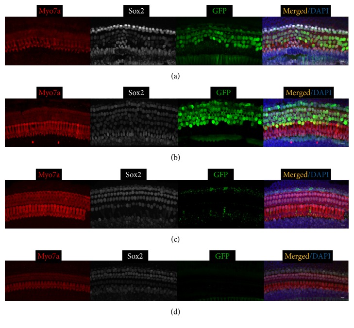
Figure 1.
Ad-Cre-GFP-Baylor transduces supporting cells when injected into mouse cochlear at P0. Representative confocal images of whole-mount fluorescent immunolabeling of cochlea injected at P0 to illustrate the basal (a), middle (b), and apical turns (c), as compared to the contralateral uninjected middle turn of cochlea (d). Myo7a labels hair cells, and Sox2 labels supporting cells. Ad-Cre-GFP-Baylor mainly transduces supporting cells in basal and middle turns. Scale bars: 10 μm.

