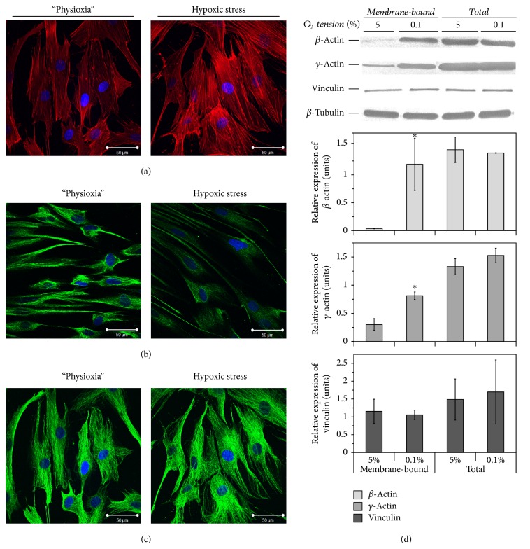
Figure 4.
The effects of hypoxic stress on cytoskeleton organisation of tissue O2-adapted adipose tissue-derived mesenchymal stromal cells (ASCs). ((a)-(c)) Representative images of phalloidin staining (a) and immunofluorescent labelling of ASCs for vimentin (b) and β-tubulin (c) before and after hypoxic stress (0.1% O2, 24 h) (n = 3). Scale bar is 50 μm. (d) Expression of β-, γ-actin, β-tubulin, and vinculin in total and membrane protein extracts obtained from ASCs before and after short-term hypoxic stress (mean + standard deviation, ∗ p < 0.05, n = 3).

