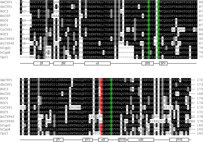
Figure 1. Multiple sequence alignment of deduced amino acid sequence of GmCYP1 with CYPs from other species.
Amino acid sequences of ROC3, BnCYP, GhCYP1, CcCYP1, ROC6, ROC1, ROC5, Cpr1, hCYP-D, hCYP-A, AtCYP40, AtCYP63 and GmCYP1 (refer to Table S1 for accession numbers) were aligned by ClustalW, and imported into BOXSHADE 3.21 for shading. Identical amino acids are shown in the dark box and similar amino acids are indicated by the grey box. Amino acid residues involved in PPIase activity (R55, F60 and H126) (Zydowsky et al.32) and CsA binding (W121) (Liu et al.33; Zydowsky et al.32) are highlighted with green and red, respectively. Secondary structure is shown below the alignment. The relative positions of amino acids indicated for PPIase activity and CsA binding sites, and the secondary structure features are based on hCYP-A (Kallen et al., 1991).

