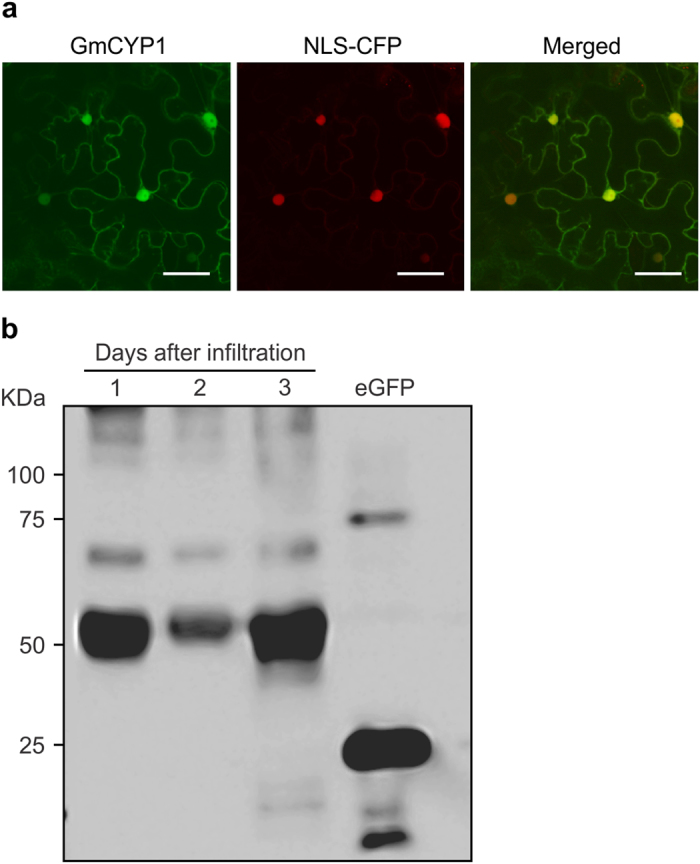
Figure 3. Subcellular localization of GmCYP1.

A. tumefaciens GV3101 carrying the plasmids with GmCYP1-YFP and nuclear localizing CFP (NLS-CFP) constructs were co-infiltrated into N. benthamiana leaves and visualized by confocal microscopy. Expression of (a) GmCYP1-YFP, (b) NLS-CFP, (c) images of A and B merged to confirm nuclear localization of GmCYP1. Scale bars indicate 50 μm. (d) Western blot analysis of translational fusion of GmCYP1-YFP proteins. GmCYP1-YFP was transiently expressed in N. benthamiana leaves for 1, 2 or 3 days, and protein accumulation was measured. Proteins (30 μg) were separated on a SDS-PAGE and transferred to PVDF membrane by electroblotting. GmCYP1-YFP protein was detected by sequential incubation of the blot with anti-GFP antibody and anti-mouse IgG conjugated with HRP, followed by chemiluminescent reaction. eGFP is shown as a positive control.
