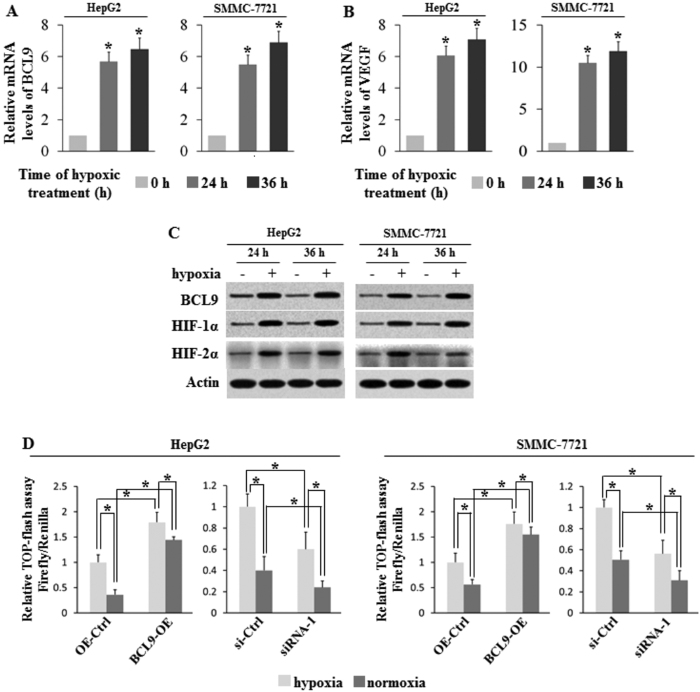
Figure 3. Hypoxia induces BCL9 expression in HCC cells.
Human HCC cell lines HepG2 and SMMC-7721 cells were cultured under the hypoxic condition for the indicated time periods. (A) The mRNA expression levels of BCL9 in these cells were determined by Taqman real-time PCR and normalized with actin. (B) The mRNA expression levels of VEGF in these cells were determined as a positive control. (C) The BCL9, HIF-1α and HIF-2α protein levels were determined by Western-blot assays. (D) Significant increase in TOPflash activity was observed under hypoxia treatment. Further, the TOPflash activity was significantly increased when BCL9 overexpressed while decreased when BCL9 knocked down. Data are presented as mean ± SD (n = 3).*p < 0.01, Student’s t-test.

