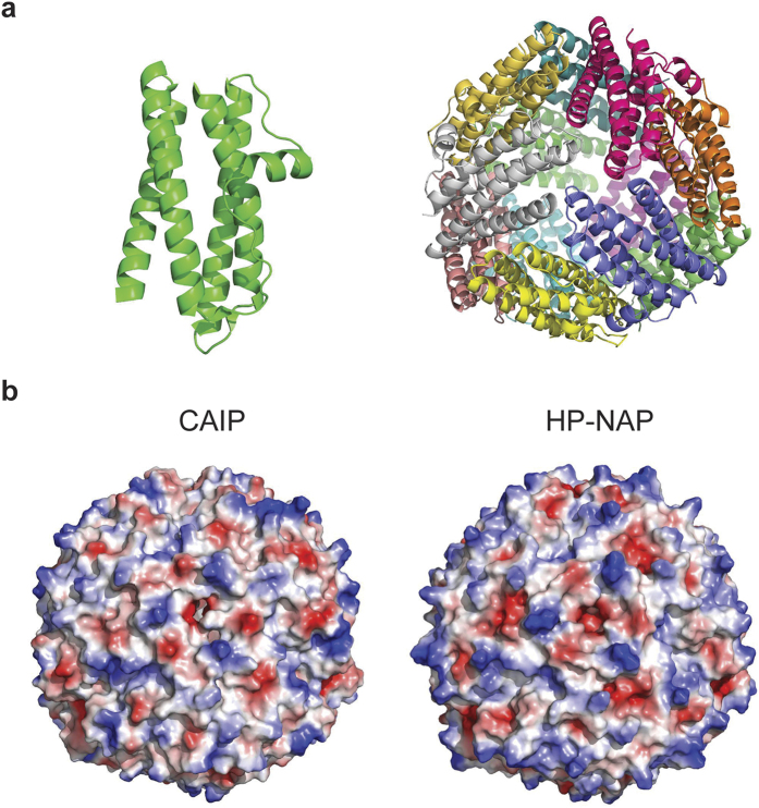
Figure 2. CAIP crystal structure.
(a) Cartoon view of CAIP monomer (left) and dodecamer (right). Each subunit is shown in a different color. One of the three-fold molecular axes is in the center of the structure, perpendicular to the plane of the paper. (b) Qualitative surface electrostatic potential of CAIP (left) and HP-NAP (right). Positive charges are in blue, negative in red. A different charge distribution on the surface of the two proteins is evident.

