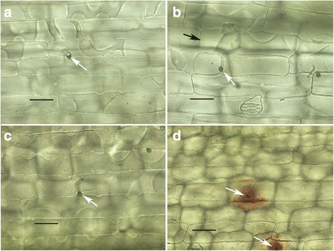
Fig. 3.

a CgSl1 on maize sheath, 48 hpi. Cell beneath appressorium (white arrow) plasmolyzes normally; b CgSl1 on maize sheath, small penetration hypha (white arrow) 48 hpi. Adjacent cell (black arrow) plasmolyzes normally. Cell containing penetration hyphae appears granulated, plasma membrane visible but appears abnormal; c M1.001 on sorghum sheath, 24 hpi, cells beneath appressoria (white arrow) still plasmolyze; d M1.001 on sorghum sheath, 48 hpi. No plasmolysis evident in any of the cells in the vicinity of the appressoria (white arrow). Scale bars equal to 50 μm
