
Fig. 1.
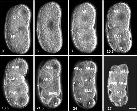
Time-lapse DIC micrographs of H. glycines early embryo development. Individual eggs were observed every half hour until the eight-cell stage. The time in hours (lower left corner) was recorded for each of the cell divisions. Cell labels are based on Caenorhabditis elegans and inferred based on positional homology in other nematode species [10, 14, 25, 27]. Scale, 15 μm