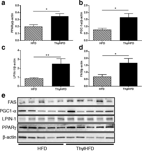
Fig. 6.

a-e Quantification of Western blot analysis of PPARγ a, PGC1-α b, LPIN-1 (Fig. c), and FAS d in WAT (n = 5 animals/group). β-actin was used for normalization. Western blot images shown in e. Bars represent median and interquartile range. Statistical difference of p < 0.05 was considered significant (*p < 0.05, **p < 0.01)
