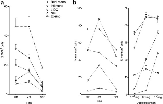
Fig. 2.

Comparison of endocytic ability of myeloid and dendritic subsets. The ability of cells to endocytose antigen was measured by uptake of OVA-FITC and mannan-FITC. Spleens were collected for analysis at the same time, and splenocytes prepared by lysis of red blood cells with enrichment for dendritic and myeloid cells via T and B cell depletion. Cells were stained with antibodies to identify L-DC and myeloid subsets as shown in Table 1. Uptake of antigen was assessed in terms of % FITC staining cells. C57BL/6 J mice were given: a OVA-FITC at 1, 3, and 6 h prior to euthanasia for spleen collection (intravenously; 1 mg per mouse). Data reflect mean ± SE (n = 4); b mannan-FITC (intravenously; 0.5 mg per mouse) at 1, 3 and 6 h prior to euthanasia for spleen collection. Single mice only were analysed tin a pilot study to determibe optimal time of 3 h used in a subsequent dose response experiment. Data reflect mean ± SE (n = 2). Control mice were given PBS
