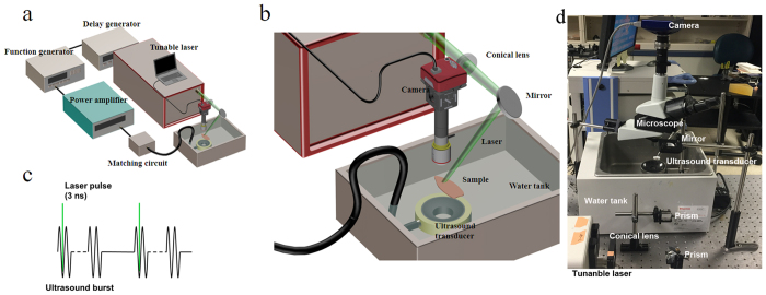
Figure 1. System schematic.
(a,b) The PUT system consisted of a laser system and a therapeutic ultrasound system. A function generator was trigged by the laser system to generate burst signals at 1 MHz and 10% duty cycle with 10-Hz pulse repetition rate. The generated bursts were amplified by a 50-dB radio frequency amplifier before being sent to the therapeutic ultrasound transducer. Either an optical microscope or a PeriCam PSI System was employed for real-time monitoring of microcirculation. (c) Each laser pulse was delivered to the sample to overlay the beginning phase of each ultrasound burst. (d) Photograph of the system.

