


Figure 2.
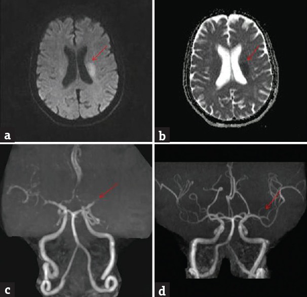
Brain magnetic resonance imaging diffusion-weighted imaging shows bright signal in the left middle cerebral artery territory (a) (red arrow); apparent diffusion coefficient shows corresponding dark signal (b) (red arrow); magnetic resonance angiography at admission shows nonvisualization of M2 (c) (red arrow); magnetic resonance angiography at 48 h postthrombolysis shows recanalization (d) (red arrow)