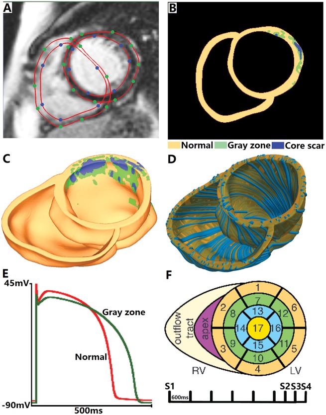
Figure 1.
Virtual-heart arrhythmia risk predictor methodology. Contrast-enhanced cardiac MRI stack with landmark points and splines delineating the endocardial and epicardial surfaces (A), and the resulting ventricular segmentation into normal tissue, grey zone, and core scar (B). High-resolution ventricular structure model (C) with estimated fibre orientations (D). Action potential (E) for non-infarcted tissue (red) and grey zone (green). Virtual-heart arrhythmia risk predictor pacing sites (F).

