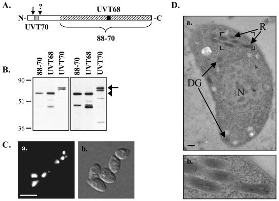
FIG. 3.
Western blot analysis and immunolocalization using ROP4-specific antibodies. (A) Schematic representation of regions recognized by the ROP4-specific antibodies. Hatched region, C-terminal 424 aa of ROP4 used as the immunogen for mouse MAb 88-70; gray and black boxes, synthetic peptides used to prepare rabbit polyclonal antibodies UVT70 and UVT68, respectively; arrow, predicted signal peptide cleavage site; arrowhead, approximate site of prodomain cleavage (see text). (B) Total RH parasite extracts were resolved by SDS-PAGE under either reducing (left) or nonreducing (right) conditions and Western blotted with MAb 88-70, UVT68, or UVT70. Arrow, pro-ROP4 (68 kDa), recognized by UVT70; arrowhead, mature ROP4 (60 kDa), recognized by MAb 88-70 and UVT68. On longer exposures, pro-ROP4 (68 kDa) was also recognized by MAb 88-70 and UVT68 (data not shown). Numbers on left indicate molecular masses in kilodaltons. (C) Indirect immunofluorescence microscopy with MAb 88-70 of methanol-fixed extracellular parasites (a) and the corresponding differential interference contrast image (b). Bar, 10 μm. (D) Immunoelectron microscopy using polyclonal antibody UVT68. ROP4 localizes specifically to the rhoptries (R), as is evident from the distribution of the 10-nm immunogold particles. N, nucleus; DG, dense granules. Bar, 200 nm. Panel b shows an enlargement of the boxed region in panel a.

