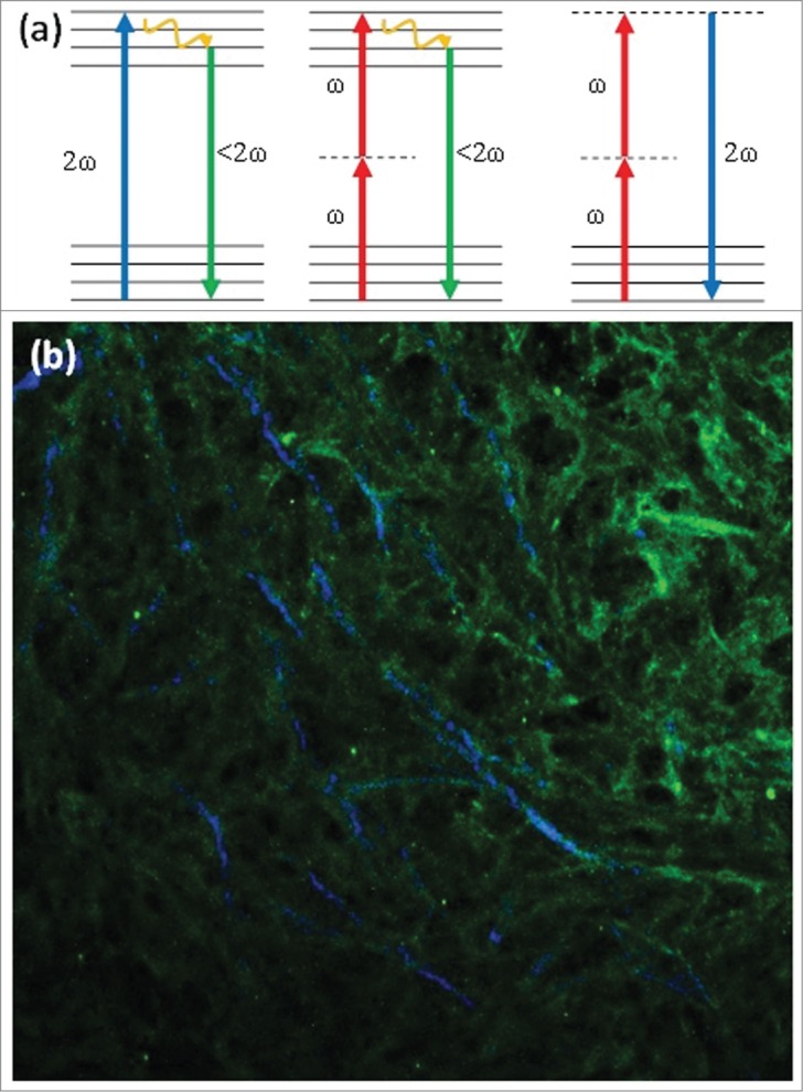
Figure 1.

(a) Jablonski diagram of (from left to right) one-photon excited fluorescence, two-photon excited fluorescence and Second Harmonic Generation, depicting the differences in excitation processes between these 3 optical processes. (b) Sample image of Type I collagen antibody staining imaged with TPEF (green) overlapped with SHG imaging of collagen fibers (blue). This image demonstrates that SHG is produced by type I collagen, but not all type I collagen produces a significant SHG signal.
