
Figure 1.
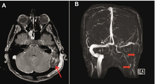
Patient MRA obtained on HD1 of first admission. A) T1-weighted precontrast imaging demonstrating a 3×4 cm area of high signal intensity extending posteriorly from the left mastoid, consistent with mastoid abscess versus osteomyelitis. B) MRV demonstrating sigmoid sinus and jugular thrombosis.