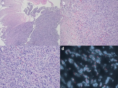
Figure 3.

The Hematoxylin and Eosin (H&E) section of the tumor cells demonstrated the proliferation of spindle cells eroding the overlying squamous mucosa (a, H&E 4×). Focal necrosis is noted (b, H&E 10×). The spindle cells showed marked cytological atypia and brisk mitoses (c, H&E 20×). FISH study on the paraffin block demonstrated CPM gene amplification (orange) in tumor cells (d, green labeled the probe for chromosome 12 centromere; orange labeled the probe for CPM gene at 12q15; aqua labeled the probe for chromosome 4 centromere as internal control).
