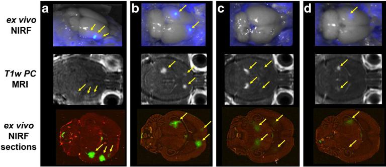
Fig. 4.
NIRF imaging demonstrates specific delivery to brain. The optical GCPII/PSMA-targeted agent YC-27 was delivered intravenously without a, b or with c, d a blocking dose of the non-fluorescent competitive agent ZJ-43. Both in situ (top) and post-transverse slice (bottom) fluorescent imaging demonstrated adequate penetration of the optical tracer to the CNS (yellow arrows), in close correlation with BBB opening as visualized by post-contrast MRI (middle, yellow arrows). Specific uptake by the brain GCPII target sites is denoted by the marked reduction of signal with ZJ-43 (Color figure online).

