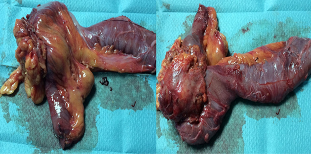
Figure 3.

Histological examination of the colonic lesion (left panel) and small intestine lesion (right panel) showing a malignant epithelial neoplasm with complex glandular architecture and intraluminal necrosis; findings typical of colonic adenocarcinoma (A and E). Both lesions share the same immunohistochemical reactivity pattern of colonic adenocarcinoma: diffuse and strong positive staining for anti-CDX2 (B and F); positive staining for anti-CK20 (C and G); and negative staining for anti-CK7 (D and H). (Hematoxylin-eosin, magnification x40 (A and E); immune-stains for CDX2 (B and F), CK20 (C and G) and CK7 (D and H), magnification x40).
