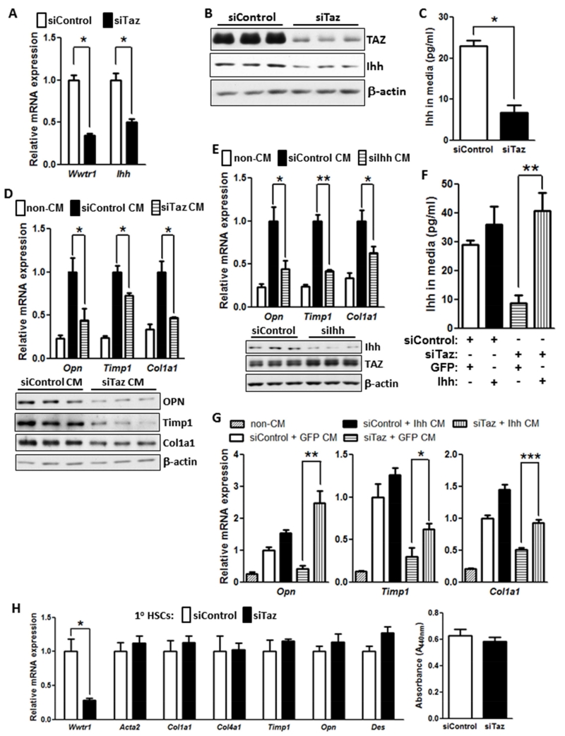
Figure 6. TAZ-Induced Hepatocyte Ihh Increases the Expression of Fibrosis-Related Genes in Hepatic Stellate Cells.
(A) Expression of Wwtr1 and Ihh mRNA in control and TAZ-silenced AML12 cells (*p < 0.0003; mean ± SEM; n=3).
(B) Immunoblot of TAZ and Ihh in control and TAZ-silenced AML12 cells.
(C) Ihh concentrations, assayed by ELISA, in the media of control and TAZ-silenced AML12 cells (*p < 0.003; mean ± SEM; n=3).
(D) Primary hepatic stellate cells (HSCs) were incubated for 72 h with conditioned medium (CM) obtained from control or Taz-silenced AML12 cells or with medium not exposed to cells (non-CM). The HSCs were then assayed for Opn, Timp1, and Col1a1 mRNA (upper panel; *p < 0.05; mean ± SEM; n=4) and the respective proteins by immunoblot (lower panel).
(E) HSCs were incubated for 72 h with non-CM or CM obtained from control or Ihh-silenced AML12 cells and then assayed for Opn, Timp1, and Col1a1 mRNA (*p < 0.04; **p < 0.0001, mean ± SEM; n=4). Immunoblot of Ihh and TAZ in siIhh-treated and control AML12 cells is shown below the graph.
(F) Control or Taz-silenced AML12 cells that were transduced with a plasmid encoding Ihh or control GFP. Aliquots of the four sets of conditioned medium were assayed for Ihh by ELISA (*p < 0.002; mean ± SEM; n=3).
(G) HSCs were incubated with conditioned media from the 4 sets of cells in (F) or with non-CM and then assayed for Opn, Timp1, and Col1a1 mRNA (*p < 0.05; **p < 0.004, ***p < 0.0004, mean ± SEM; n=4). Note that bars 2 and 3 for Opn and Col1a1 are significantly different at p < 0.05.
(H) Primary HSCs were activated by culturing for 72 h in medium containing 10% FBS and then treated with siControl or siTaz duplex, followed by culturing in the same medium for an additional 48 h. The cells were then assayed for Wwtr1, Acta2, Col1a1, Col4a1, Timp1, Opn, and Des mRNAs or cell proliferation. For the proliferation assay, the cells were synchronized by overnight culture in medium containing 0.2% FBS and then cultured an additional 48 h in medium containing 10% FBS. The cells were incubated with WST-1, and absorbance at 440 nm was measured to assess cell number.

