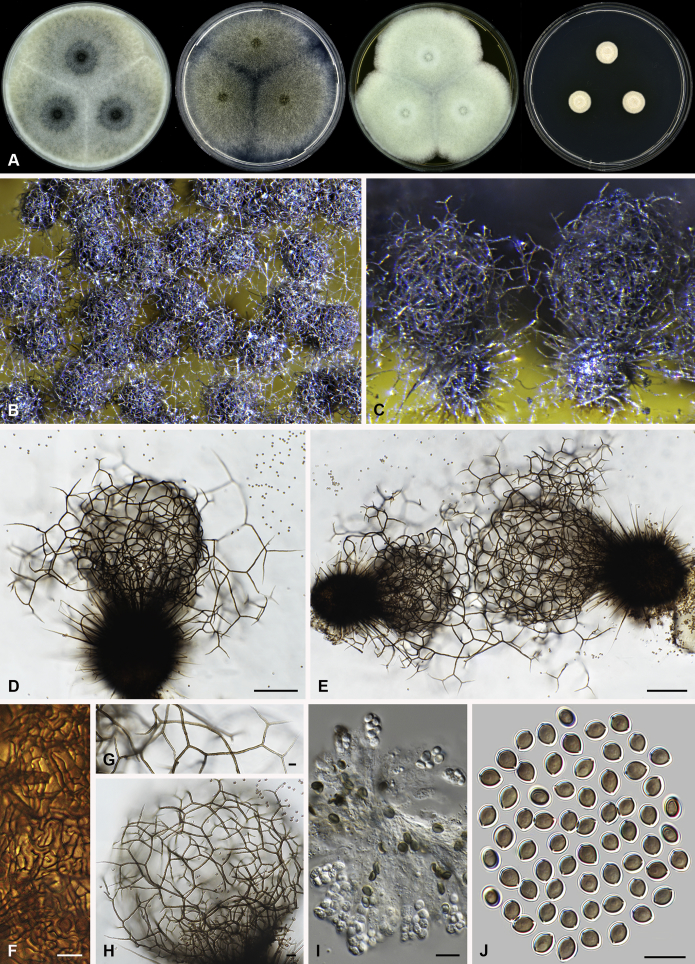
Fig. 38.
Dichotomopilus variostiolatus (DTO 319-A2). A. Colonies from left to right on OA, PCA, MEA and DG18 after 7 d incubation. B. Mature ascomata on OA, top view; C. Mature ascomata on OA, side view. D–E. Ascomata mounted in lactic acid. F. Structure of ascomatal wall in surface view. G. Upper part of a terminal ascomatal hair. H. Net structure formed by terminal ascomatal hairs. I. Asci. J. Ascospores. Scale bars: D–E = 100 μm; H= 20 μm; F–G, I–J = 10 μm.

