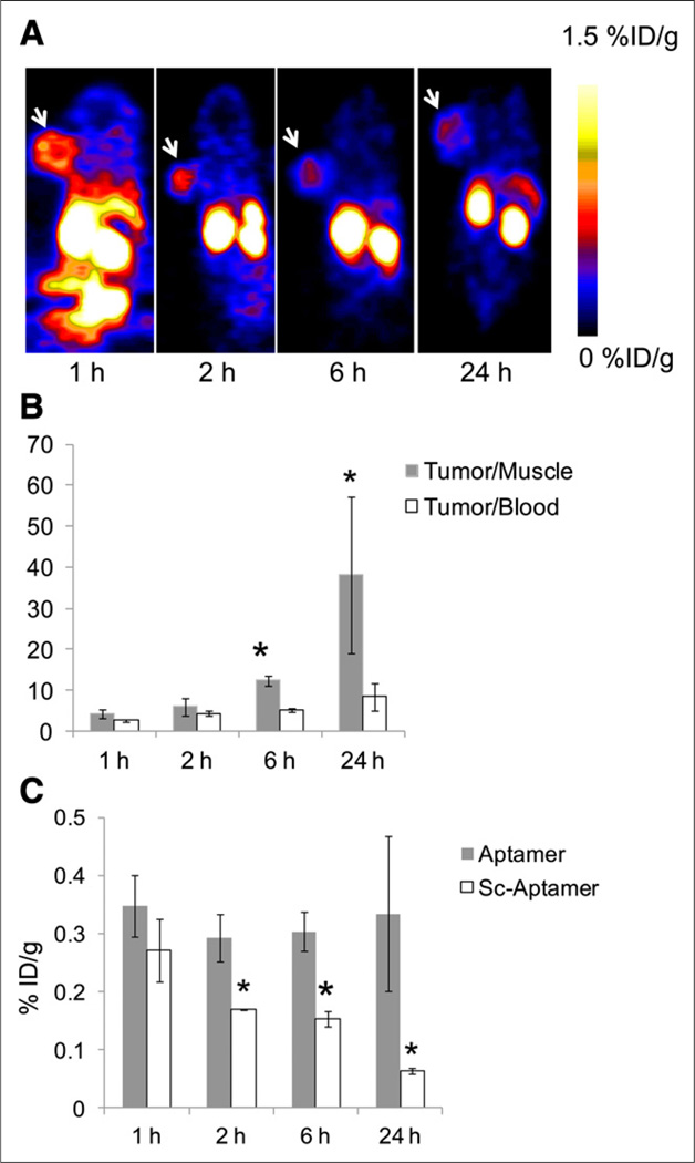
FIGURE 5.
(A) Representative coronal PET images of mice at 1, 2, 6, and 24 h after injection of 64Cu-NOTA-tenascin-C aptamer. White arrow indicates tumor. (B) T/M and T/B ratios of 64Cu-NOTA-tenascin-C aptamer over time. Results are presented as average of 5 mice ± SD. *Significance between 6 and 24 h and 1 and 2 h. (C) Comparison of tumor uptake between 64Cu-NOTA-tenascin-C aptamer and 64Cu-NOTA-Sc aptamer (n = 5/group). *Significance between aptamer and Sc aptamer tumor uptake.

