
Figure 3. Transplantation of small mitochondria into femoral arteries by intravenous administration.
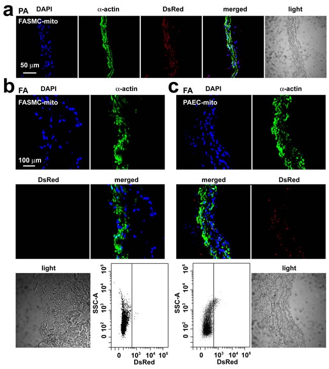
a. Immunohistochemical stainings of pulmonary artery (PA) in rats after an intravenous injection of DsRed-labeled mitochondria prepared from femoral artery smooth muscle cells (FASMC-mito, width ~296 nm) showing DAPI (blue), SMC marker α-actin (green), DsRed (red), overlap of the above three (merged) and light field. b.-c. Immunohistochemical stainings of femoral artery (FA) in rats after an intravenous injection of DsRed-labeled FASMC-mito (width ~296 nm, b) or DsRed-labeled mitochondria prepared from pulmonary artery endothelial cells (PAEC-mito, width ≤ 150 nm, c) showing DAPI (blue), SMC marker α-actin (green), DsRed (red), overlap of the above three (merged) and light field, and the representative flow cytometry for the separation of DsRed-labeled exogenous mitochondria from PA. The mitochondria isolated from FA of rats without mitochondrial injection were used as a negative control to set up voltages of side scatter (SSC) for flow cytometry analysis. Each represents stainings of 9 artery segments from 3 animals.