
Figure 2. urantide inhibits IRF3 expression and activation in LPS-stimulated KCs.
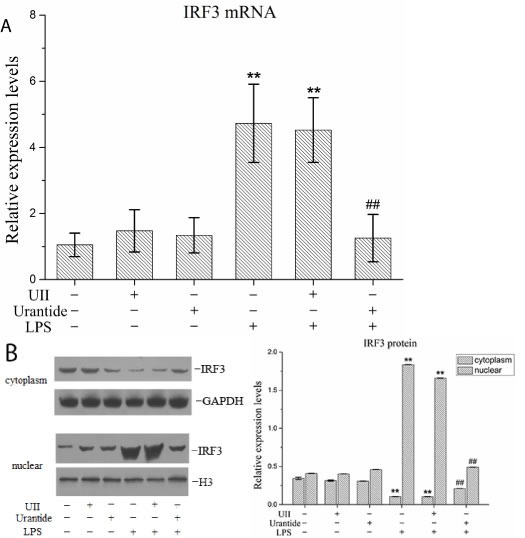
KCs were treated with UII or urantide 0.5 h before LPS stimulation. A. IRF3 mRNA expression in KCs using real-time PCR. Relative expression levels of IRF3 mRNA were detected in KCs after normalization to GAPDH through real-time PCR. Data represent means ± SD (n = 6). B. IRF3 protein expression in the nuclear and cytoplasm of KCs using Western blot. Left panel shows representative pictures of Western blot, and right shows relative levels of nuclear and cytoplasm IRF3 protein after normalization to H3. Data represent means ± SD (n = 6). *P<0.05 and **P<0.01 versus control cells [UII(-)urantide(-)LPS(-)]; #P<0.05 and ##P<0.01 versus LPS-stimulated cells [UII(-)urantide(-)LPS(+)].