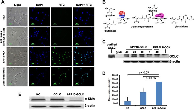
Figure 7. Efficient transduction of short peptide or protein in fibrosis related HSC-T6 cells in vitro.

(A)Apoposis induced by after hPP10-KLA incubation in HSC-T6 cells, TUNEL assay was performed for apoptosis detection. (B) Schematic diagram of the glutathione biosynthesis biological process. (C) After treating with different dosage of hPP10-GCLC fusion protein on HSC-T6 cells for 1 h, intracellular localization of hPP10-GCLC were detected by Western blotting, purified GCLC was used as postive control. (D) Intracellular glutathione (GSH) was detected using GSH-Glo™ Glutathione Assay. (E) After incubated with hPP10-GCLC on HSC-T6 cells, alpha-SMA expression was analyzed by Western blotting.
